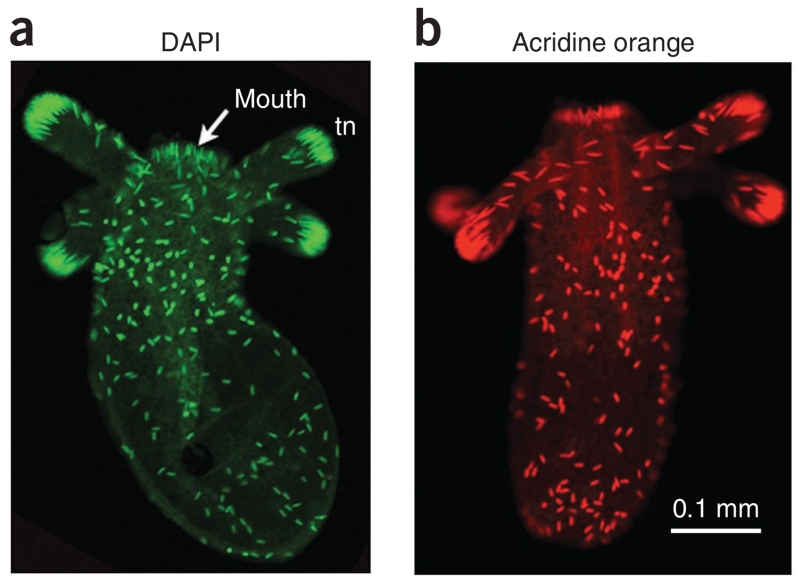
Figure 4.
Example of cnidocyte staining. Nematostella juvenile polyps were fixed and stained under calcium-free conditions. (a) DAPI staining of the poly-γ-glutamate of cnidocyte capsules was detected in a green (521 nm) emission channel. (b) AO also stains cnidocyte capsules, and its fluorescence was detected in a red (615 nm) emission channel. In both stains, cnidocytes are primarily detected in the ectoderm of the body column.

