Figure 1.
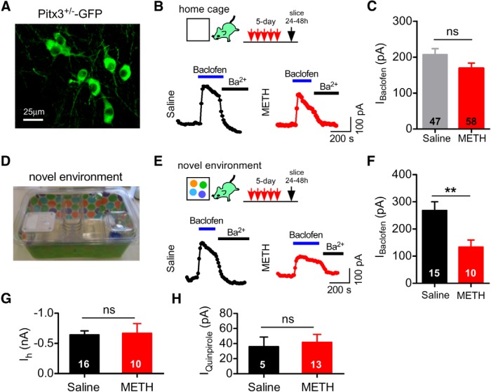
Attenuation of IBaclofen in VTA DA neurons after 5 d METH injections in a novel environment. A, Live fluorescent image of GFP+ DA neurons in the VTA of a Pitx3+/−-GFP mouse. B, Examples of maximal (300 μm) IBaclofen recorded from VTA DA neurons 24–48 h after 5 d of intraperitoneal injections of either saline (0.9%) or METH (2 mg/kg in 0.9% saline) in the home cage. Application of 1 mm BaCl2 inhibits IBaclofen. C, Mean IBaclofen recorded 24–48 h after 5 d saline or 5 d METH injected in the home cage (ns, p > 0.05 Student's t test). D, Photograph of the novel cage environment with different bedding and brightly colored paper. E, Examples of IBaclofen recorded from VTA DA neurons 24–48 h after 5 d injections of saline or METH in a novel environment. F, Mean IBaclofen in VTA DA neurons 24–48 h after 5 d injections of METH or saline in a novel environment (**p < 0.01, Student's t test). G, No difference in mean Ih between 5 d saline- or METH-injected mice. H, No change in mean IQuinpirole (30 μm) with 5 d METH compared with 5 d saline in a novel environment.

