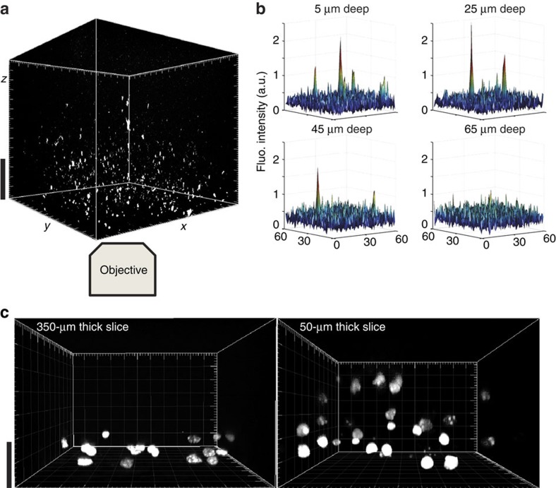
Figure 4. Imaging penetration depth analysis of the detection of single QD in acute brain slices.
(a) 3D reconstruction of z-stack acquired with spinning disk confocal microscopy in an acute brain slice of a rat injected with QD (scale bar, 20 μm). The imaged QD can be detected in live tissue at least up to depths of 45 μm (b), as exemplified with surface view of the image pixel levels. Membrane permeability assays were performed with PI staining, showing a layer of damaged cells in 350 μm slices and much more damage in 50 μm thick slices (c) (scale bar, 20 μm).

