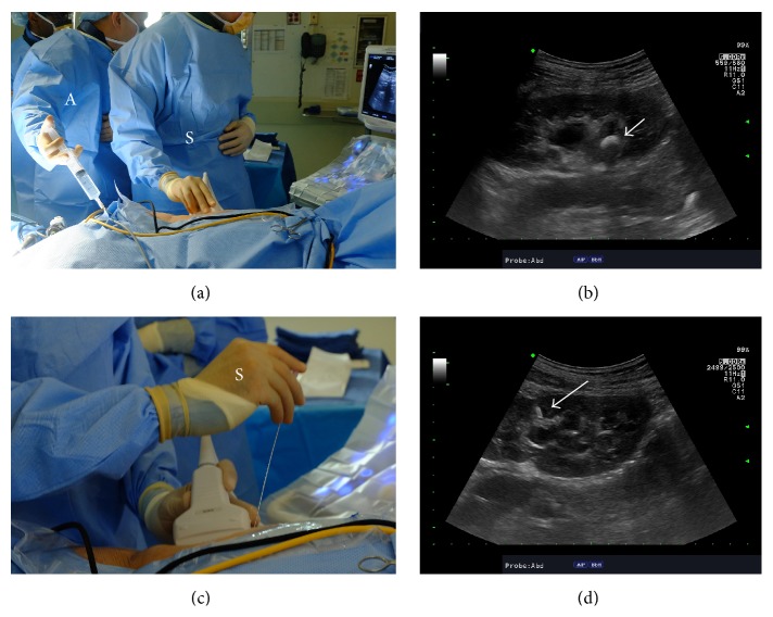
Figure 1.
Establishing renal access using ultrasound guidance. (a) The operative surgeon (S) holds the ultrasound probe and the assistant (A) holds a syringe attached to ureteral catheter for normal saline infusion if needed. (b) Ultrasonographic image of the kidney along its longitudinal axis demonstrating the stone in the renal pelvis (white arrow) within a mildly hydronephrotic collecting system. (c) During the needle insertion, the operative surgeon (S) holds both the ultrasound probe and the needle to perform the puncture. For hand positioning, the nondominant hand holds the ultrasound probe while the dominant hand holds the needle. (d) The needle can be visualized (white arrow) entering the collecting system through upper pole calyx in this case.

