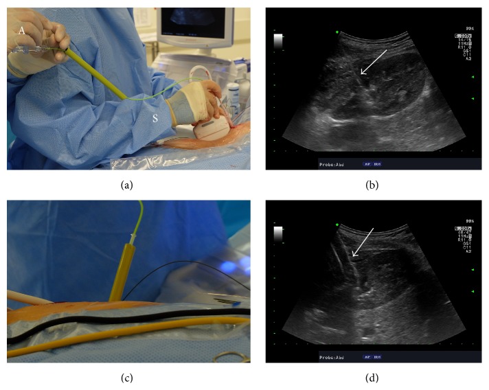
Figure 3.
Tract dilation with high-pressure balloon under ultrasound guidance. (a) The deflated balloon dilator is inserted over a working wire and for this step, the operative surgeon (S) controls the ultrasound probe and distal end of the balloon while the assistant (A) controls the wire on the proximal end of the balloon dilator. (b) Ultrasonographic image of the kidney along its longitudinal axis demonstrates that the tip of the deflated balloon dilator (white arrow) is difficult to visualize and differentiate from the wire. (c) The sheath has been inserted over the inflated balloon dilator that is subsequently withdrawn. (d) The inflated balloon (white arrow) can be readily seen with ultrasound imaging.

