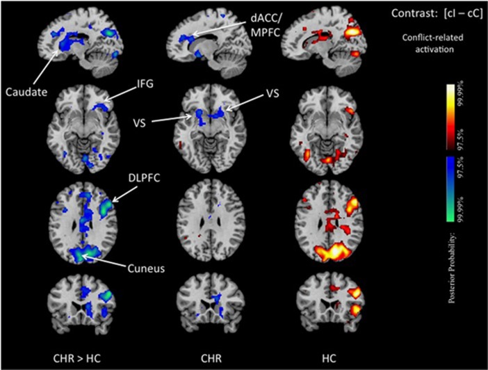
Figure 2.
Group differences in conflict-related activation. We show posterior probability maps, which we thresholded at 97.5%. Increases in signal during correct responses to cI relative to cC trials are shown in red and decreases in blue. This group comparison shows decreased conflict-related activations in CHR participants compared with healthy controls in the dorsal caudate, IFG, ventral striatum, the DLPFC, the dACC/MPFC and at the border between cuneus and precuneus (left column). Within-group effects are displayed separately for CHR patient (middle column) and healthy controls (right column) and confirm that the reduced conflict-related activations detected in the between-group comparison were driven by positive activations in controls and, in CHR participants, either by significant deactivations in the dACC and ventral striatum or, for the other regions, by a lack of detectable conflict-related activations. The reduced conflict-related activations found in the CHR group represented areas where functional activity associated with the conflict-free condition (cC) was higher than that associated with the conflict-dependent condition (cI). dACC/MPFC, dorsal anterior cingulate/medial prefrontal cortex; DLPFC, dorsolateral prefrontal cortex; IFG, inferior frontal gyrus; VS, ventral striatum.

