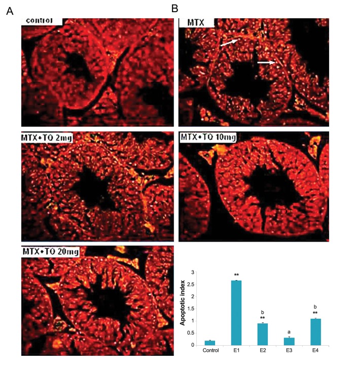
Fig.1.
The apoptosis inducing effect of methotrexate (MTX) (20 mg/kg) and different doses of thymoquinone (TQ) on testis of mice. A. Images by a fluorescent microscopy indicating terminal deoxynucleotidyl transferase dUTP nick end labeling (TUNEL) staining of mice testicular sections that are counterstained with propidium iodide (PI). Apoptotic cells show bright fluorescence nuclei indicated by arrowheads (magnifications: ×160) and B. Percent of TUNEL positive cells (AI). The mice were grouped as Control, MTX (E1), MTX+TQ 2 mg/kg (E2), MTX+TQ 10 mg/kg (E3), MTX+TQ 20 mg/kg (E4). **; P<0.001 compare to control group, a; P <0.01 and b; P<0.05 compare to MTX group.

