
Figure 2. Structure of the epimerase part of GNE.
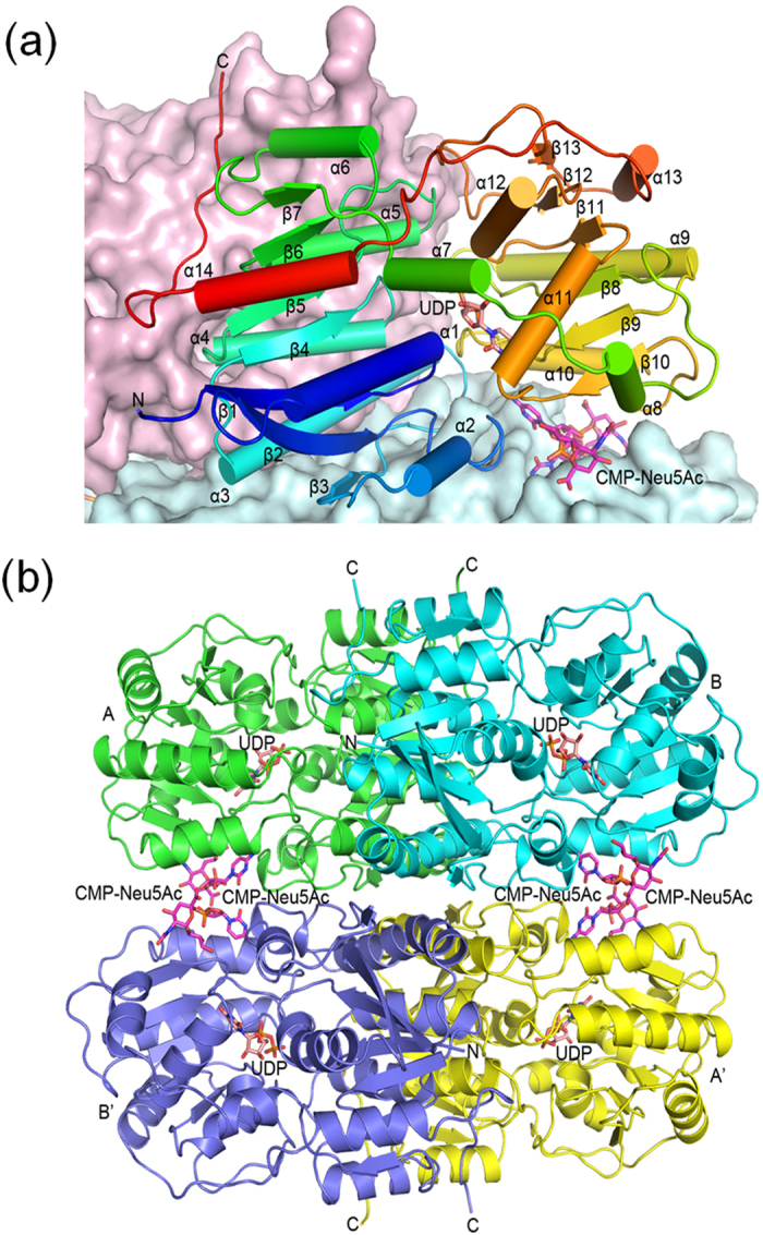
(a) A monomer is shown as a ribbons diagram, with cylinders and arrows representing α-helices and β-strands. The model is colored from N-terminus to C-terminus by a spectrum from blue to red. Also shown are surface representations of the other monomers in the same dimer (colored light pink) and the tetramer-forming dimer (light blue). The bound UDP in the active site is depicted as a pink stick model. Two CMP-Neu5Ac molecules at the dimer-dimer interface are colored magenta. (b) The tetramer is shown as a ribbons diagram where the subunits are in four different colors. Monomers A and B form a dimer and monomers A’ and B’ form another. The stick models of UDP and CMP-Neu5Ac are colored pink and magenta.