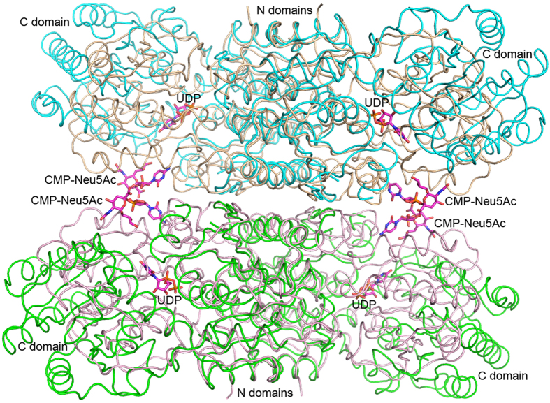
Figure 3. Open and closed conformations of the GNE tetramer.
The tetramer is shown as a worm tracing diagram, with the two dimers colored light pink and salmon. Two copies of the dimer of nonhydrolyzing epimerase from M. jannaschii (PDB 4NEQ) are superimposed on GNE by using the N domain as in supplementary Fig. S1b. These are colored cyan and green.

