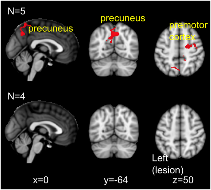
Figure 3. Resting state connectivity with left ipsilesional motor cortex.

First row: regions with increased resting state connectivity with left motor cortex at post 1 relative to pre-intervention. Second row: no changes in resting state connectivity between baseline and pre-intervention time points. Results presented using cluster thresholding at z > 2.5, p < 0.05 corrected.
