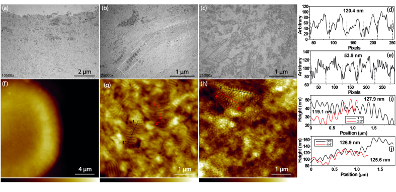
Figure 3. The TEM and AFM images of the wide-spaced collagen of FECD-DMs.
(a) The wide-spaced collagen distributed on the posterior of the DM; (b) Different types of collagen in the FECD-DMs; (c) The morphology formed by the wide-spaced collagen together with other fibrillar collagen structures. (d,e) The line profiles of two types of collagens labelled in two white dash boxes in (b), which indicate different periodicities of different collagen types; (f) AFM image of a guttae; (g) The zoom in image on the guttea in (f); (h) AFM image near the guttae, the Z range in f is from 0 μm to 3.0 μm while that of in g and h are from 0 nm to 164.6 nm; (i,j) The line profiles of four color lines in (g,h), respectively.

