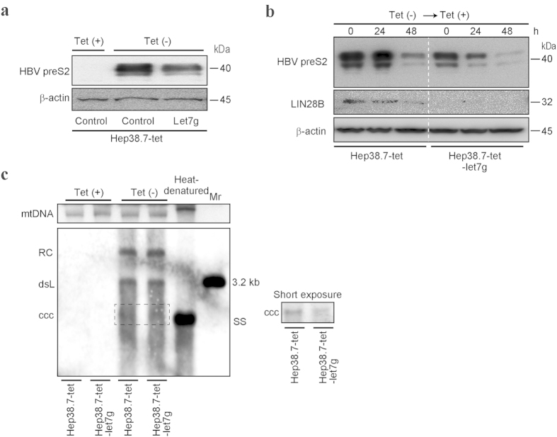
Figure 4. Let-7 g decreases HBV preS2 protein expression.
(a) Hep38.7-tet cells were cultured long-term with or without tetracycline (Tet). HBV preS2 protein expression was determined by Western blotting. Let-7 g overexpression suppressed preS2 protein levels. Representative results from three independent experiments are shown. (b) Hep38.7-tet and stable let-7 g-overexpressing Hep38.7-tet cells were cultured without tetracycline (Tet), and then HBV transcription was shut off by adding tetracycline (Tet). PreS2 protein levels were determined at the indicated time points by Western blotting. LIN28B expression levels were also evaluated. Representative results from three independent experiments are shown. (c) HBV cccDNA levels in the indicated cells with or without Tet were determined by Southern blotting. DNA extracted by the Hirt method was applied. Mitochondrial DNA (mtDNA) was used as the loading control. Heat-denatured DNA indicated the disappearance of double-stranded DNA. Double-stranded linear full-length HBV DNA (dsL) was used as a 3.2 kb marker (Mr). RC, relaxed circular DNA. ccc, cccDNA. SS, single stranded DNA. A short-exposure image is also shown to identify cccDNA (right). Representative results from three independent experiments are shown.

