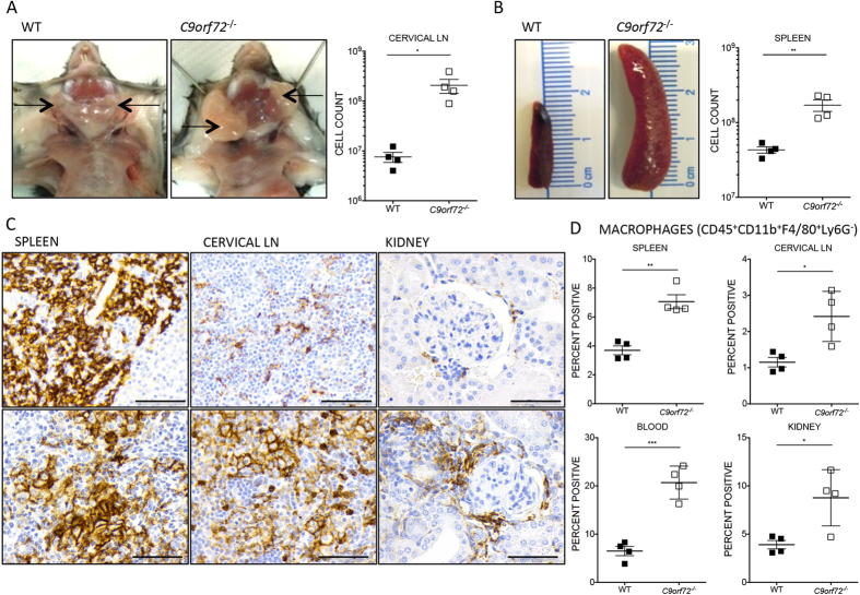
Figure 1. C9orf72−/− mice develop lymphadenopathy and splenomegaly, and display infiltration of F4/80+ cells by IHC and FACS Analysis.
(A,B) Representative pictures of gross cervical LN enlargement and splenomegaly observed in C9orf72−/−in comparison to age-matched WT control. Significantly increased cell counts obtained via FACS analysis correspond to lymphadenopathy and splenomegaly observed grossly. (C) The expanded cell populations infiltrating the red pulp of the spleen and surrounding lymphoid follicles of the cervical LN stained positive by IHC for mouse macrophage marker F4/80. Periglomerular infiltrates observed in C9orf72−/− kidneys are also largely positive for F4/80 macrophage lineage marker. Sections shown are females, 37 week old C9orf72−/− and 40 week old WT (D) FACS analysis confirmed H&E and IHC findings by showing increased percentages of CD11b+F4/80+Ly6G− macrophages in kidney, spleen, cervical LN, and blood (30–35 week old female, n = 4 per genotype). (A–D) Data are shown as mean ± s.e.m (*P ≤ 0.05, **P ≤ 0.01 and ***P ≤ 0.001 by unpaired Students t-test). (C) Scale bar represents 50 μm, original magnification, ×600.

