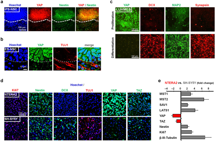
Figure 1. Neural YAP expression decreases upon neuronal differentiation.
(a) Immunocytochemistry of YAP (red) and nestin (green) in human iPS cell-derived neural cultures (IMR90-4-DL1 iPS cells; scale bar: 100 μm). (b) Immunocytochemistry of YAP (green) and neuronal β- III-tubulin (TUJ1; red) of human ES cell-derived neural cultures (H9 ES cells; confocal microscopy; dashed line in a,b indicates neuroepithelial boundary; scale bar: 10 μm). (c) Immunocytochemistry of YAP (green) and neuronal markers doublecortin (DCX; red), MAP2 (green) and Synapsin (red) under proliferation vs. differentiation conditions of an immortalized human neural cell line (LUHMES). (d) Comparative immunocytochemistry for markers of proliferation (Ki67, Nestin), markers of neuronal differentiation (DCX, TUJ1) and YAP and TAZ expression in the human embryonal carcinoma NTERA2 cell line versus SH-SY5Y neuroblastoma cells grown under proliferative conditions. (e) Quantitative RT- PCR analysis comparing SH-SY5Y (grey bars) versus NTERA2 (red bars) mRNA expression fold changes for inhibitory upstream members of the Hippo signaling pathway (MST1/2, SAV1, LATS1), the transcriptional co-activators YAP and TAZ and neural stemness (Nestin, Ki67) and differentiation (β- III-tubulin) markers (n = 3 independent experimental repeats; error bars indicating SEM).

