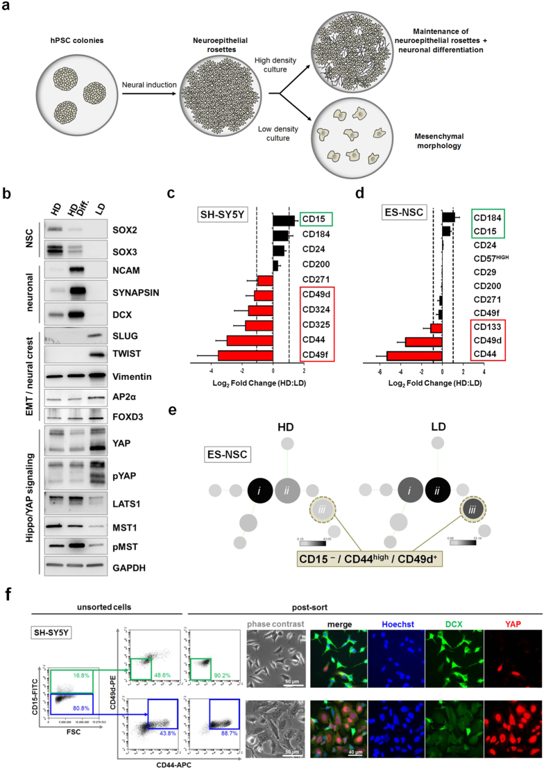
Figure 2. Low density culture conditions promote a neural crest cell phenotype and a CD44+ /CD49d+ YAP-positive subpopulation.
(a) Schematic for the generation of PSC-derived neural cultures. (b) Western blot analysis of low density-conditioned neuronal cultures (LD) in comparison to high density-conditioned neuroepithelial stem cells (HD) and neuronally differentiated neuroepithelial stem cells (HD Diff.) derived from human ES cells. Expression of the EMT markers SLUG and TWIST are seen in the LD cultures only, together with a relative increase in the mesenchymal marker Vimentin and the neural crest markers AP2α and FOXD3, together with YAP. (c,d) Fold change of surface marker expression between high and low density conditions (c) in SH-SY5Y and (d) in human ES cell-derived neuronal cultures, determined by flow cytometric analysis. Bars represent average ± SD (n = 2 independent experiments). (e) Unbiased clustering algorithm (SPADE) showing cell frequency changes between high and low density conditions in main clusters (i,ii,iii) (f) Fluorescence-activated cell sorting (FACS) enrichment of YAP-positive cells from heterogeneous SH-SY5Y culture using a CD15–/CD44+/CD49d+ code (far right dot plot panels showing post-FACS reanalysis; scale bars: 50 μm and 40 μm, as indicated).

