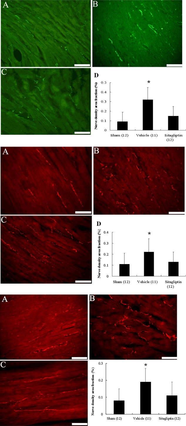
Figure 2. Upper, immunofluorescent staining for tyrosine hydroxylase from the remote regions (magnification 400×).

Tyrosine hydroxylase-positive nerve fibres are located between myofibrils and are oriented longitudinal direction as that of the myofibrils. Middle, immunofluorescent staining for growth-associated protein 43 from the remote regions (magnification 400×). Lower, immunofluorescent staining for neurofilament from the remote regions (magnification 400×). (A) Sham; (B) infarction treated with vehicle; (C) infarction treated with sitagliptin. Bar=50 μm. nerve density area fraction (%) at the remote zone. Each column and bar represents mean ± S.D. *P<0.05, compared with sham and sitagliptin.
