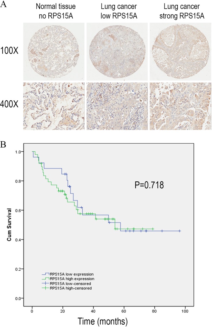
Figure 1. Immunostaining of RPS15A with tissue microarray.

Immunostaining of RPS15A in lung adenocarcinoma and adjacent normal tissues with tissue microarray. (A) Three representative cases with different expression status of RPS15A, ranging from negative, mild and strong expression were taken at 100 × and 400 × magnification in lung cancer and normal tissues. (B) Kaplan-Meier survival analysis of overall prognosis between patients with higher RPS15A expression and patients with low RPS15A expression. Log-rank test was used to statistically calculate the difference.
