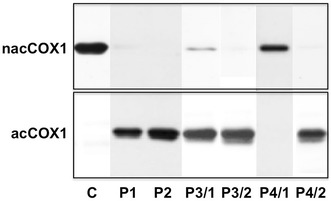
Fig. 1.

Detection of acetylated COX1 (acCOX1, lower panel) and non-acetylated COX1 (nacCOX1, upper panel) in platelet lysate of non-treated controls (C) and patients on aspirin treatment (P1–P4). Monoclonal antibodies specific to the acetylated and non-acetylated forms of COX1 were used in Western blotting detection system. In patients P3 and P4 non-compliance was presumed and the test was repeated after a 2 weeks period of compliance (P3/2 and P4/2). Below the band representing COX-1 another low intensity band cross-reacting with both antibodies is also apparent. It very likely represents COX-1 isoform 2, a 37 amino acids shorter transcript variant (http://www.ncbi.nlm.nih.gov/nuccore/18104968)
