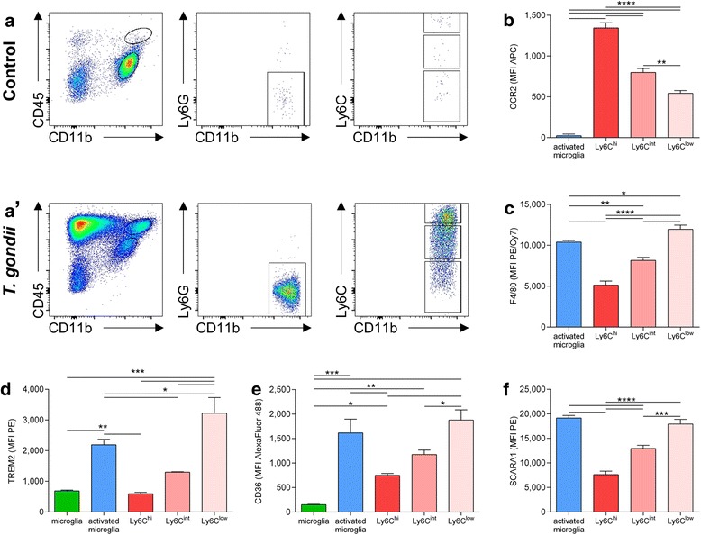
Fig. 3.

Myeloid-derived mononuclear cells are recruited to the brain upon T. gondii infection and express phagocytosis related surface molecules. Mononuclear cells were isolated from 5xFAD mouse brains and subjected to flowcytometric analysis. a, a’ Representative pseudocolor plots are shown for (a) non-infected and (a’) infected 5xFAD animals and demonstrate the recruitment of CD45hiCD11bhiLy6GnegLy6C+ cells to the brain upon T. gondii infection. After gating cells by their forward and side scatter properties, excluding doublets and dead cells (not shown), we used CD45 and CD11b expression to discriminate between resting microglia (a, bottom elliptic gate) or activated microglia (a’, bottom elliptic gate), respectively, and myeloid cells (a’, top elliptic gate). From myeloid cells, Ly6G+ neutrophils were excluded and the Ly6C expression of the remaining CD11bhiLy6G− cells was used to gate Ly6Chi, Ly6Cint and Ly6Clow mononuclear cells. b–f We compared the surface expression of CCR2, F4/80, TREM2, CD36, and SCARA1 between resting microglia, activated microglia and myeloid-derived mononuclear cell subsets. The median fluorescence intensity (MFI) for each marker and population is displayed as mean + SEM. Significance levels (p values) determined by Fisher’s LSD test are indicated. *p ≤ 0.05, ***p ≤ 0.001, ****p ≤ 0.0001
