
Fig. 6.
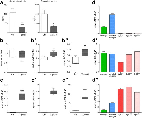
T. gondii infection reduces small and large Aβ aggregates and enhances mRNA expression of the Aβ degrading enzymes IDE and MMP9 as well as immunoproteasomal subunits. a Aβ42 in the carbonate soluble (monomeric and small oligomeric Aβ42 aggregates) and guanidine soluble fractions (large Aβ42 aggregates) of whole-brain homogenates was measured by ELISA. Data are presented as mean + SEM. b, b’, b” Expression of neprilysin (NEP), matrix metalloproteinase 9 (MMP9) and insulysin (IDE) in the brain was measured by RT-PCR in non-infected (n = 5) and T. gondii infected (n = 7) 5xFAD mice. Data are presented as fold-change over non-infected 5xFAD mice in box and whisker graphs. c, c’, c” Expression of the immunoproteasome subunits LMP2, LMP7 and MECL-1 in the brain was measured by RT-PCR in non-infected (n = 5) and T. gondii infected (n = 7) 5xFAD mice. Data are presented as fold change over non-infected 5xFAD mice in box and whisker graphs. d, d’, d” Expression of the MMP9, IDE and LMP7 by innate immune cells isolated and sorted from the brains of non-infected and T. gondii infected C57BL/6 mice was measured by RT-PCR. Each sample consists of pooled cells from six animals and was measured in triplicates. Significance levels (p values) determined by unpaired Student’s t test or Fisher’s LSD test are indicated. *p ≤ 0.05, **p ≤ 0.01, ****p ≤ 0.0001