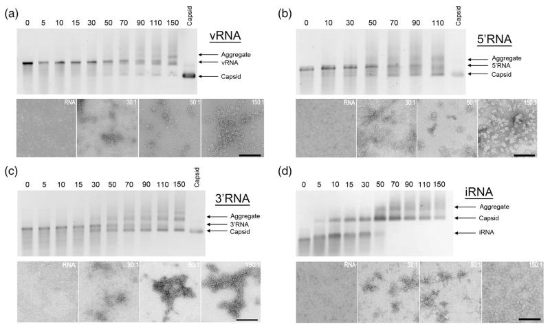
Fig. 2.
Gel mobility and electron microscopy assays of MS2 capsid assembly. Assembly reactions with (a) vRNA, (b) 5′RNA, (c) 3′RNA, and (d) iRNA. The top half of (a – d) shows a native agarose gel of capsid assembly reactions induced with the respective RNA. The number above each lane indicates the CP2:RNA stoichiometry; i.e. coat protein dimer:RNA, of the assembly reaction in that lane. The migration positions of the RNA fragment used in each panel, the recombinant MS2 capsid and aggregated material are indicated. Selected reactions were negatively stained with 2% (w/v) uranyl acetate and imaged by electron microscopy. The scale bars represent 200 nm.

