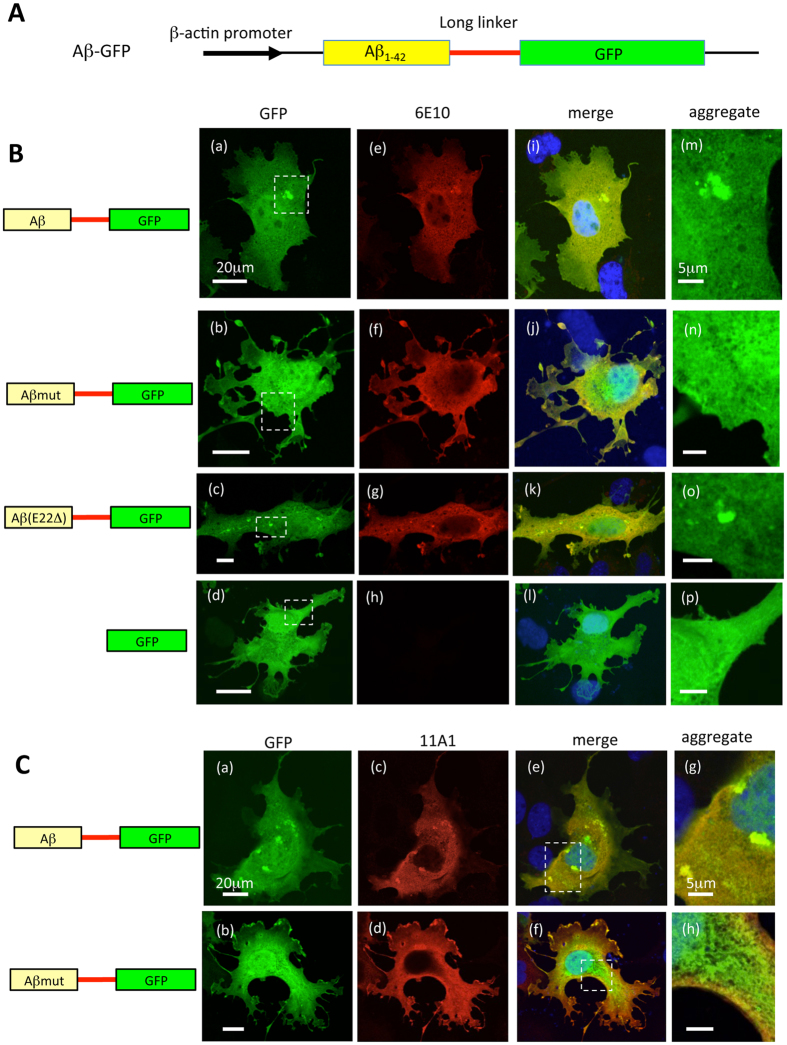
Figure 1. Representative images of COS7 cells transfected with various Aβ-GFP DNA constructs.
(A) Basic structure of genes encoding fusion protein containing Aβ1-42 fused to GFP with a long-linker sequence (14 amino acids). (B) COS7 cells were transfected with plasmids encoding Aβ-GFP (a) Aβmut-GFP (b) Aβ (E22Δ)-GFP (c), or GFP (d). To confirm the expression of Aβ proteins, transfected cells were immunostained with the 6E10 antibody (e–h). Merged images with GFP are shown in (i–l). The regions within the dotted rectangles in (a–d) are enlarged in (m–p). Aggregated Aβ proteins (dotted localizations) were observed in Aβ-GFP and Aβ (E22Δ)-GFP transfected cells, however, the Aβmut-GFP proteins did not form detectable aggregates in cells. Scale bars: 20 μm (a–d) 5 μm (m–p). (C) Immunostaining of COS7 cells expressing the Aβ-GFP or Aβmut-GFP fusion proteins with the 11A1 antibody. Merged images showed that almost all the Aβ-GFP fusion protein was labeled with the11A1 antibody, indicating that the Aβ-GFP fusion protein formed oligomers. In contrast, the Aβmut-GFP was only partially labeled with the11A1 antibody. Scale bars: 20 μm (a–f) 5 μm (g,h).

