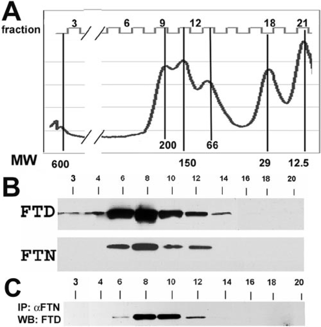
Figure 1.
Gel filtration chromatography of proteins extracted with HEPES buffer from E15 embryonic CE tissue. (A) Calibration curve showing the molecular weight makers in kilodaltons, and the corresponding numbers of the fractions collected and subsequently analyzed for ferritoid (FTD) and ferritin (FTN) by denaturing SDS-PAGE, followed by Western blot analysis. (B) Western blot analysis of the fractions probed for FTD and FTN after denaturing SDS-PAGE. (C) Immunoprecipitation of the fractions with anti–FTN antibody followed by Western blot analysis for FTD.

