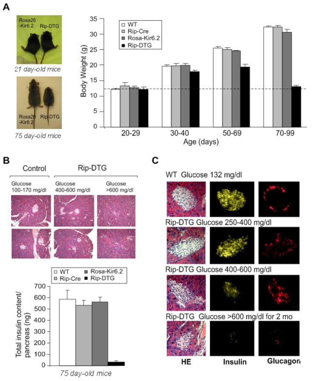
Figure 2. Growth retardation accompanied by dramatic changes in islet morphology and hormone content in diabetic Rip-DTG animals in late stages of diabetes.
(A) (left) Photographs of Rip-DTG mice and single transgenic littermates at 21 days and 75 days. (right). Body weight as a function of age for Rip-DTG, WT and single genotype littermates (n = 12–15 in each case). (B) (top) Low magnification (20x) pancreatic sections from Rip-DTG and control animals stained with hematoxylin-eosin and (lower) pancreatic insulin content as a function of age for Rip-DTG, WT and single genotype littermates (mean ± SEM, n = 4–8 mice in each case). (C) Hematoxylin-eosin staining (left), insulin (middle) and glucagon (right) immunostaining of pancreatic paraffin sections from WT control (top) and Rip-DTG mice at early (middle) and late stages of disease (below).

