Fig. 1.

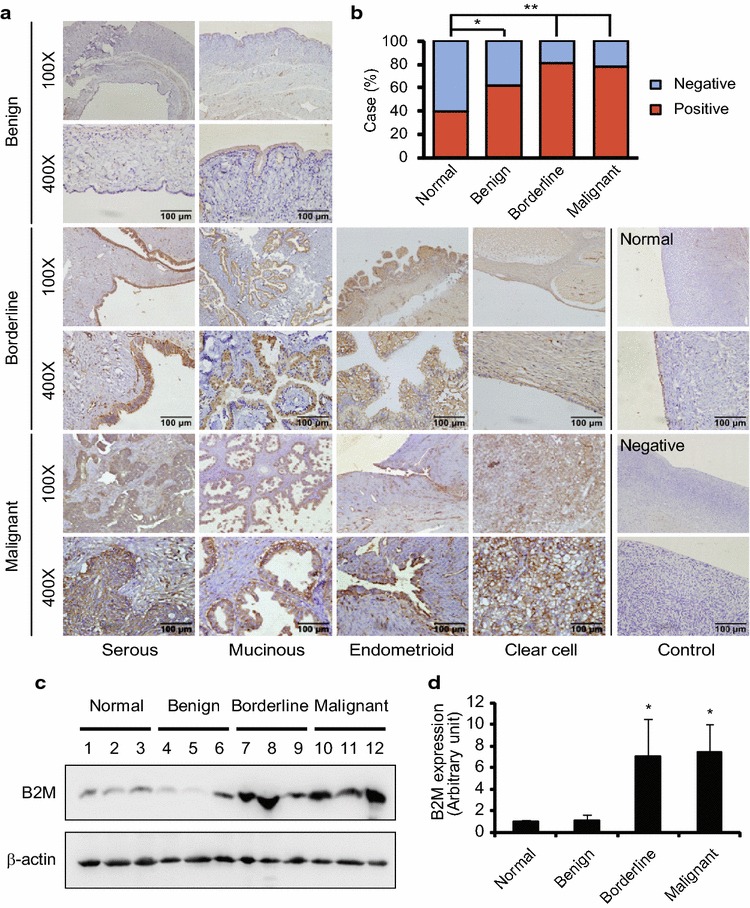
B2M protein expression in human ovarian tissues. a Immunohistochemical staining of B2M protein in human epithelia-type ovarian tumours. A brown color in epithelial cells is considered as a positive staining. Negative control without first antibody is performed in the normal ovarian tissue. Representative images of B2M expression in serous, mucinous, endometrioid and clear cell tumours and the normal ovarian tissue are shown. Original magnification × 100 and × 400. Scale bar 100 µm. b The case rate of B2M positive and negative. Positive vs. negative: 16/24 in control without tumour (40 cases), 24/14 in benign tumour (38 cases), 31/7 in borderline tumour (38 cases) and 25/7 in malignant tumour (32 cases). For comparison between two groups, χ2 test was applied. c Detection of B2M expression in the normal ovarian tissues (case #1–3) and serous benign (case #4–6), borderline (case #7–9) and malignant (case # 10–12) tumours by Western blot. d Semi-quantitative analysis after densitometry on the gels of (c). Benign, benign tumour; Borderline, borderline tumour; Malignant, malignant tumour; Normal, normal ovarian tissue. *P < 0.05; **P < 0.01
