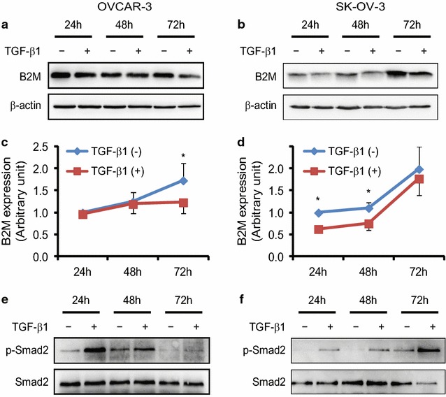
Fig. 4.

Effect of TGF-β1 on the expression of B2M protein in ovarian cancer cell lines. In a time-course study, OVCAR-3 (a) and SK-OV-3 (b) cells were treated with 10 ng/ml of TGF-β1 for 24, 48 and 72 h, respectively. Equal amounts of total protein were subjected to SDS-PAGE and transferred to a PVDF membrane. Specific signal was detected by Western blot analysis using a specific antibody against B2M or β-actin. c, d The graphs show the quantitative analysis of the gels from OVCAR-3 and SK-OV-3 cells, respectively, after densitometry (both n = 3). β-actin was served as a loading control. *P < 0.05. Phospho-Smad2 (p-Smad2) and total Smad2 were used as indicators for the TGF-β signaling pathway existed in those cells. p-Smad2 was increase upon TGF-β1 stimulation in OVCAR-3 (e) and SK-OV-3 (f) cells
