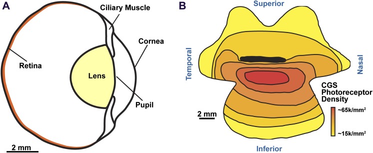
Fig. 1.
A cross-sectional schematic of 13LGS ocular anatomy (A). The horizontal optic nerve head (ONH) lies approximately where “retina” is indicated. Reproduced from Chou & Cullen (1984) and Sussman et al. (2011) with permission. Schematic of photoreceptor density in relation to the horizontal ONH (dark black line) of the California ground squirrel (B). The red area of highest cone density denotes the visual streak. Reproduced from Long & Fisher (1983).

