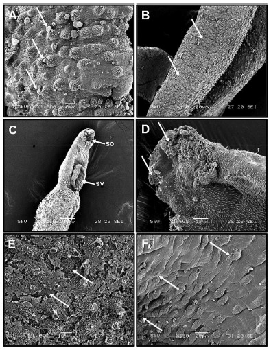
Fig. 2. (A-F). Electromicrographs of S. mansoniadult male worms of treated with different concentrations of MVEO.(A) After 24 h of MVEO (500 µg mL-1)treatment, bubble lesions were spread over the entire body of the worms (arrow). (B) Ventral portion of the adult worms ofS. mansoni after 24h of incubation with MVEO (500 µg mL-1). The loss of tubercules in some regions was observed (arrows). (C) Anterior region of the adult male worms 48 h after incubation with 250 µg mL-1 of MVEO. Destruction of the oral (os) and ventral (vs) suckers. (D) Tegument lesion severity increased (arrows) after 72 h of MVEO treatment (100 µg mL-1). (E) Tegument erosion (arrows) can be visualized at a higher magnification with no spines after 96 h of exposure to 10 µg mL-1 of MVEO. (F) Destruction of some tubercules after 120 h of incubation with 5 µg mL-1of MVEO (arrows).

