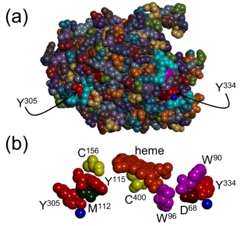
Figure 3.
(a) Space-filling structural model of the heme domain of CYP102A1 (PDB #2IJ2) highlighting the surface locations of terminal residues in pathways I (Tyr334) and II (Tyr305). (b) Space-filling model of the residues comprising CYP102A1 radical transfer pathways I and II. Blue spheres represent structurally resolved water molecules.

