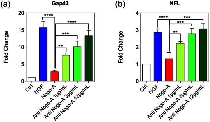
Figure 5.
Gene expression changes for neuronal markers. PC-12 cells’ mRNA expression levels of (a) Gap43 and (b) NFL, showing neural maturity during culture stimulated by NGF, and post-treated with Nogo-A peptide or three different concentrations of Nogo-A antibody. The intensities for the GAP43 and NFL genes in the Nogo-A antibody-treated groups were significantly upregulated when compared to the Nogo-A peptide group. Statistically significant differences were *p < 0.05, **p < 0.01, and ***p < 0.001 in each group with respect to the Nogo-A peptide group.

