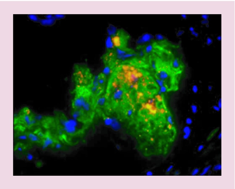
Figure 5. . Using quantum dots to image the monocyte-macrophages in atherosclerosis plaque.
The monocyte-macrophages loaded by cell penetrating quantum dots were injected to mice. Injected cells and macrophage marker CD68 are portrayed as orange and green, respectively.
Reproduced with permission from [59], © (2010) Current Atherosclerosis Reports.

