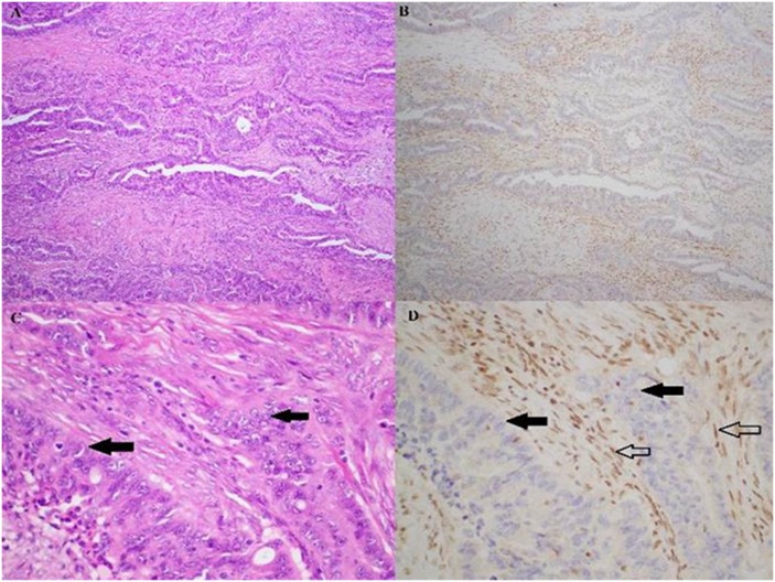
Fig 2. Haematoxylin & eosin (A, C) and BAP1 immunohistochemistry (B, D) stained sections from the sole pancreatic ductal adenocarcinoma patient who expressed loss of nuclear BAP1.
In this case, the exocrine cells (solid arrows) clearly lacked the brown BAP1 staining, and the non-neoplastic endothelial cells serve as the internal positive controls (hollow arrows). Magnifications: A, B 100x; C, D 400x.

