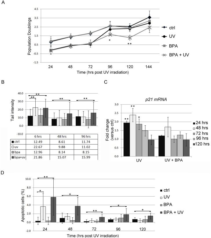
Fig 5. Analysis of FRTL-5 cell response to UV-C irradiation following long-term BPA treatment.
(A) Proliferation rate analysis of FRTL-5 cells treated for 28 days with 10−9 M BPA and then subjected to UV-C irradiation. Cells were counted every 24 hrs until 144 hrs post-irradiation. Population doubling was calculated as described in Materials and Methods. Data are reported as mean ± standard deviation of three independent experiments. (B) Quantification of DNA damage by comet assay. Data are reported as mean ± standard deviation of the tail intensity of around 100 cells analyzed for each point. (C) qRT-PCR analysis of the pattern of p21 transcript levels following UV-C irradiation. Data are reported as the ratio between p21 transcript levels in irradiated and control cells. The mean ± standard deviation of three independent experiments is reported. *p-value <0.05; **p-value <0.01. (D) Quantification of apoptotic cells by TUNEL staining. Data are reported as percentage of TUNEL positive cells per total cell number identified by DAPI staining. The results are expressed as mean ± standard deviation of several fields analyzed in three independent experiments.

