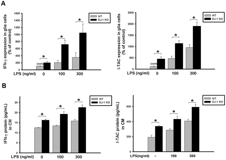
Fig 6. LPS-induced increase of IFN-γ and I-TAC is up-regulated in DJ-1 knockout mixed-glia cultures.
Primary mixed glia cultures were derived from the brain of 1-day-old WT and DJ-1 KO mice. (A) The mRNA levels of IFN-γ and I-TAC measured by quantitative real-time RT-PCR were higher in DJ-1 KO cells than WT cells following LPS administration (100 and 300 ng/ml) for 24 hours. (B) The protein levels of IFN-γ and I-TAC measured by ELISA were increased in conditioned medium (CM) of DJ-1 KO cells as compared with WT cells following application of LPS (100 and 300 ng/ml) for 48 hours. Data were normalized as percentage of the mean of basal expressional levels in WT mice (con) and presented as mean ± S.E.M. (n = 4–5 for each group) * p<0.05.

