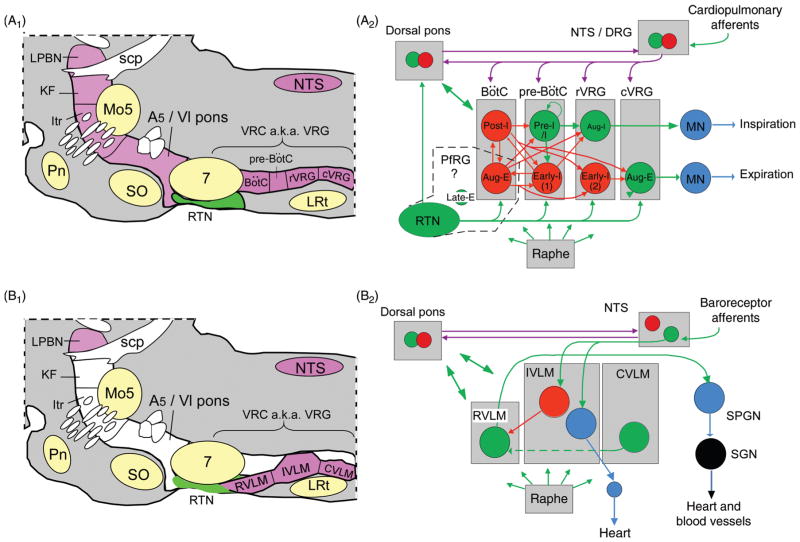
Figure 2.
Pontomedullary regions responsible for eupneic breathing and for generating the autonomic outflows to the cardiovascular system: anatomy and simplified circuitry. (A1) Parasagittal section through the pons and medulla oblongata of a rodent. The regions colored in magenta contain the principal building blocks of the respiratory pattern generator. The ventral respiratory column (VRC) contains four functional compartments aligned in rostrocaudal order (Bötzinger Complex (BötC), pre-BötC, rostral ventral respiratory group (rVRG) and caudal VRG (cVRG)). The retrotrapezoid nucleus (RTN) resides at the rostral end of the VRC under the facial motor nucleus. In this article, the term RTN refers specifically to a cluster of about 2000 CO2-activated Phox2b-ir glutamatergic neurons (in rats, 800 in mice). (A2) Minimal circuitry responsible for the generation of eupneic breathing [adapted, with permission, from Lindsey, Ryback & Smith (246)]. The drawing depicts some of the neuronal interconnections within and between the four compartments of the ventral respiratory column and a few of the connections of RTN neurons (for details see text). The parafacial respiratory group (pfRG) is a physiologically defined entity now believed to be specifically involved in the generation of active expiration (330). Its constituent neurons and their location are not yet defined. Bötzinger augmenting expiratory neurons have been included by some authors in the pfRG (327). Inhibitory (GABAergic or glycinergic) neurons are represented in red, glutamatergic neurons in green, motoneurons in blue, connections with both excitatory and inhibitory components in magenta (e.g., neurons transmitting information from arterial baroreceptors, pulmonary stretch afferents, the carotid bodies etc.). (B1) The regions colored in magenta are thought to contain the main components of the network that generates the autonomic outflows to the cardiovascular system. From an autonomic regulation standpoint, the ventrolateral medulla can be subdivided into three regions whose anatomical relationship with the respiratory compartments can be appreciated by comparing panels A1 and B1. (B2) Schematic of cardiovagal parasympathetic neurons, RVLM presympathetic neurons and connections responsible for their regulation by arterial baroreceptors. Abbreviations: aug-E, augmenting expiratory neurons; aug-I, augmenting inspiratory neurons (a.k.a. inspiratory premotor neurons); CVLM, caudal VLM; DRG, dorsal respiratory group (caudolateral portion of the NTS); early-I, early-inspiratory neurons, early-I(1) and early-I(2) are postulated to have distinct input-output functions; IVLM, intermediate VLM; Itr, intertrigeminal region; KF, Kölliker-Fuse nucleus; LPBN, lateral parabrachial nuclei; LRt, lateral reticular nucleus; Mo5, trigeminal motor nucleus; NTS, nucleus of the solitary tract; pfRG, parafacial respiratory group; Pn, pontine nuclei; post-I, postinspiratory; preI/I, preinspiratory/inspiratory (putative rhythmogenic neurons); scp, superior cerebellar peduncle; SO, superior olive; SPGN, sympathetic preganglionic neurons; SPN sympathetic (post)ganglionic neuron; VRC, ventral respiratory column.

