Figure 9.
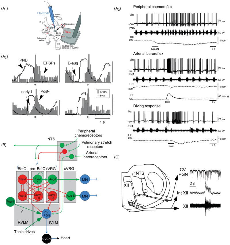
Regulation of the cardiovagal parasympathetic outflow by the respiratory pattern generator. (A1–3) [Reproduced from McAllen et al. (277), reprinted with permission.] Intracellular recordings of cardiovagal ganglionic neurons from an arterially perfused rat preparation in which the lungs are not inflated therefore reflexes from lung stretch receptors are absent (A1). The firing properties of cardiovagal preganglionic neurons (CVPGN) were inferred from the discharge pattern of the ganglionic neurons (A2) or the EPSPs recorded from these neurons (A3). Activation of the carotid bodies with cyanide or of arterial baroreceptors by raising perfusion pressure produced the expected robust excitation of cardiovagal neurons, and so did the activation of the diving reflex (A2). A3, PND-triggered histograms of EPSPs recorded in cardiovagal ganglionic neurons (CVGNs) indicate that, in this preparation, CVPGNs discharge preferentially during the postinspiratory phase. (B) Schematic interpretation based on A2–3 and C. Rat cardiovagal preganglionic neurons could be receiving inhibitory inputs from early-I and Aug-E neurons. The existence of inhibitory input during inspiration has been documented in slices as shown in C. Pulmonary stretch receptors (inactive in an arterially perfused preparation) inhibit cardiovagal preganglionic neurons, possibly via a direct input from GABAergic “pump cells” located within the NTS. Carotid body stimulation produces opposite effects on CVPGN activity depending on the intensity of the stimulus. Mild stimulation inhibits these neurons via the RPG, presumably via inhibitory inputs from early-I and post-I neurons as shown in B. Acute and intense stimulation of the carotid bodies (e.g., with cyanide) activates cardiovagal preganglionic neurons as shown in A2. This effect may be mediated by polymodal NTS neurons that also respond to noxious stimulation of the airways (pathway not represented). Post-I glutamatergic neurons are an alternative plausible source of excitation of cardiovagal preganglionic neurons and other vagal efferents with a post-I discharge pattern but the existence of such neurons has yet to be demonstrated. (Panel C was originally published in (153), and has been reproduced by permission of Oxford University Press [http://www.oxfordscholarship.com/view/10.1093/acprof:oso/9780195306637.001.0001/acprof-9780195306637]).

