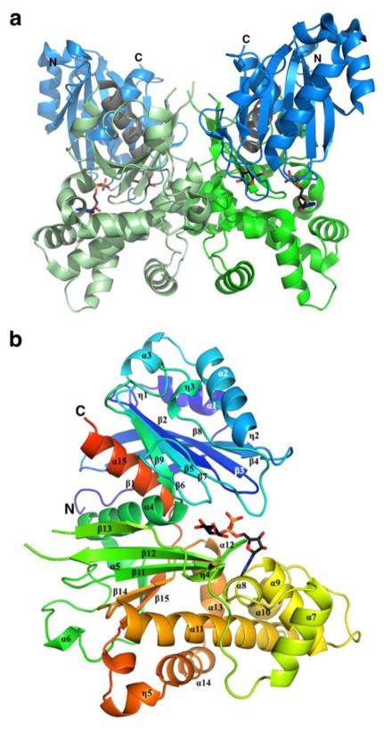
Figure 2.
Dimeric and monomeric representations of the TcGlcK-ADP-D-glucose complex (PDB entry 2Q2R) (31). (a) TcGlcK dimer with N- and C-termini indicated for each subunit and is color-coded as follows: blue for the small domain, green for the large domain, and grey for the linking segments between domains. The same colors as slightly shaded represent the other subunit. (b) TcGlcK monomer revealing α-helices, 310-helices, and β-strands in sequential order. Secondary structure elements were produced by the DSSP program from PDB entry 2Q2R, as exhibited in Figure 1 of reference (31).

