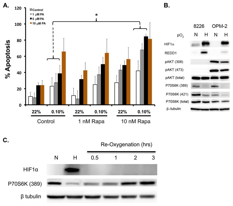
Figure 7.
Combination of rapamycin and HIF-PA can overcome resistance to hypoxia-mediated apoptosis in 8226 cells. (A) 8226 cells were cultured under normoxic (22%O2, 5% CO2) or hypoxic (0.1% O2, 5% CO2) and treated with the indicated concentrations of rapamycin, HIF-PA or both for 24 hours. At the end of the treatment, cells were harvested and % apoptosis was measured by flow cytometry for cleaved caspase 3. Values are mean ± 1 standard deviation of 3 independent experiments. Bracket * indicates significant change (P<0.05) of treatment compared to control (ANOVA). The effect of combining HIF-PA with rapamcyin on induction of apoptosis was assessed by the median effect method using Calcusyn Software Version 1.1 (Biosoft, Cambridge, United Kingdom). Combination indices (CI) values were calculated using the most conservative assumption of mutually nonexclusive drug interactions. CI values were calculated from median results of apoptosis assays. (B) Immunoblots of mTOR-related signaling proteins in 8226 and OPM-2 cells cultured under normoxic or hypoxic conditions for 24 hours. The data presented are a representative immunoblots of at least 2 independent experiments. (C) Time course of p70S6 kinase phosphorylation in OPM-2 cells following re-oxygenation. Cells were cultured under hypoxic conditions (0.1% O2, 5% CO2) for 24 hours, then cultured under normoxic conditions (22% O2, 5% CO2). Cell lysates were harvested at indicated time points and immunoblotted for HIF1α and phospho-P79S6K (389) expression. The data presented are a representative immunoblots of at least 2 independent experiments.

