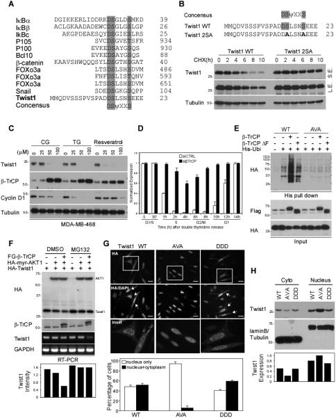
Figure 4. AKT1 induces β-TrCP-mediated Twist1 degradation.

(A) Sequence alignment of β-TrCP destruction motif. Twist1 contains one putative β-TrCP recognition motifs (DSΨXXS). D, Aspartic acid; S, Serine; Ψ, hydrophobic; X, any amino acid.
(B) Pulse-chase experiment of Twist1 WT and 2SA protein stability using cycloheximide.
(C) MDA-MB-468 cells were treated with CG, TG and resveratrol at the indicated concentration for 48 h and subjected to Western blotting with the indicated antibodies.
(D) Quantification of Twist1 expression during cell cycle progression. HeLa cells carrying siCTRL or siβ-TrCP were synchronized using double thymidine block. Protein expression was measured by Western blot and quantified by densitometer. The full figure is presented in Figure S4D.
(E) Covalently conjugated His-ubiquitin of Twist1 was pulled down by Ni2+ agarose beads under denaturing condition and analyzed by Western blot with indicated antibodies.
(F) Western blot analysis of Twist1 degradation in HEK-293T cells.
(G) Subcellular localization of Twist1 WT, Twist1 AVA and Twist1 DDD. HA-Twist1 WT, AVA and DDD were transiently expressed in HeLa cells. The localization of Twist1 variants was imaged under a fluorescent microscope.
(H) Cell fractionation analysis of Twist1 variants.
