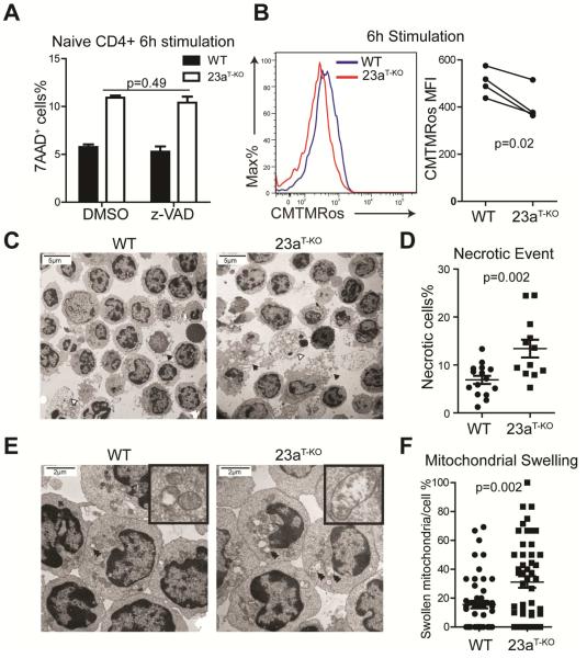
Figure 5. Deletion of Mir23a augments necrosis in CD4+ T cells at the early activation stage.
(A) Effect of caspase inhibitor z-VAD-fmk on cell death in activated CD4+ T cells from WT and Mir23af/fCd4-cre (shown in this figure as 23aT-KO) mice. These data represent three independent experiments. (B) Mitochondrial potential during early priming in naïve WT and miR-23a-deficient CD4+ T cells. The right panel is the quantification of 4 independent experiments. (C) Transmission electron microscopy (TEM) for activated WT and miR-23a-deficient CD4+ T cells (2050×). Black arrows mark the cells undergoing necrosis and white arrows mark the apoptotic cells. (D) Quantification of necrosis rates for activated T cells. The data represent 15 1250× fields for the WT group and 12 1250× fields for the Mir23af/fCd4-cre group. (E) TEM for activated WT and miR-23a-deficient CD4+ T cells (5600×). Double arrows mark the swollen and ruptured mitochondria. (F) Quantification of mitochondrial swelling in activated CD4+ T cells. The data represent 50 individual cells for each genotype. See also Figure S4.

