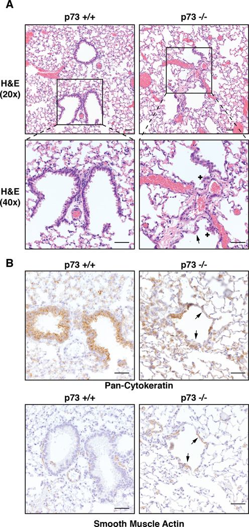
Figure 2. p73−/− Mice Exhibit Severe Airway Phenotypes Including Hyperplasia and Epithelial Loss.
(A) Representative H&E images of the terminal airways in 18 month old p73+/+ and p73−/− mice. The p73−/− mice exhibit areas of epithelial loss (arrow) and small nodules of hyperplastic epithelium (+). (B) Immunohistochemistry (IHC) staining of pan-cytokeratin and α-SMA of above mice. Arrows in panel B indicate areas of epithelial loss as well as hypertrophy and hyperplasia of the smooth muscle (scale bar= 50 µm).
See also Figure S2.

