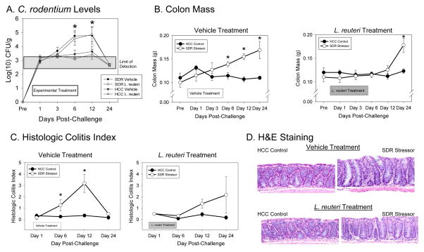
Figure 1.
Exposure to the SDR stressor significantly changed C. rodentium colonization, pathogen-induced increases in colon mass, and histopathology. Mice were exposed to the SDR stressor during oral challenge with 3–5 × 10⁶ CFU of C. rodentium. A). Exposure to the SDR stressor led to increased levels of C. rodentium that could be cultured from the stool during the first 24 days post-challenge. Treatment with L. reuteri did not affect the stressor-induced increase in pathogen challenge. B). Colon mass was significantly increased in mice exposed to the SDR stressor during oral challenge with C. rodentium. This increase did not occur in mice treated with L. reuteri until Day 24 post-challenge. C). Colonic histopathology was significantly increased in mice exposed to the SDR stressor during oral challenge with C. rodentium. This increase was not evident in mice treated with L. reuteri. D). Representative images of H&E stained colonic sections (magnification = 20X). In all cases, the data are the mean ± S.E. * indicate p<.05 vs non-stressed HCC control mice at the same time point.

