Figure 1.
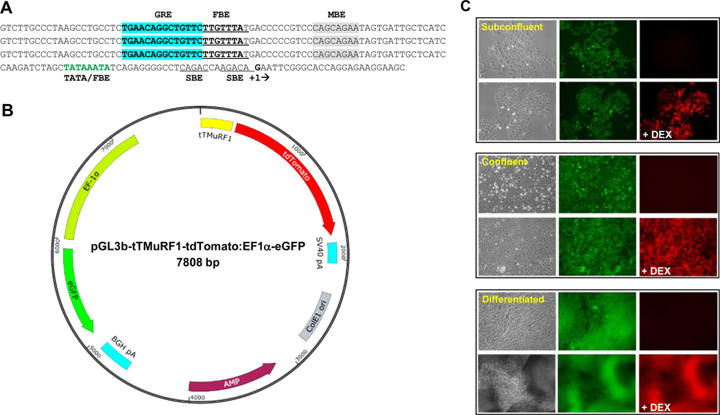
Sequence of the tTMuRF1 promoter, schematic representation of pGL3b-tTMuRF1-tdT:EF1a-eGFP vector, and in vitro testing of To3B cells. (A) Triple tandem (tT) repeat of the sequence of a putative GRE-FBE (in bold-type and with GRE highlighted in blue) along with flanking sequence from the human MuRF1 proximal promoter region fused to its core promoter, i.e., the indicated TATA box that overlaps with another FBE (in green-type) with transcription start-site boxed (+1). SMAD binding elements (SBE, double underline) and putative myogenin binding element in gray highlight are also indicated. (B) Simplified schematic map of our dual reporter vector with tTMuRF1 promoter, tdTomato, simian virus 40 polyadenylation signal (SV40 polyA), EF1α promoter, eGFP, bovine growth hormone (BGH) polyA, ampicillin (AMP) resistance, and E. coli origin of replication (ColE1 ori) sequences mapped to their respective positions and orientations within the pGL3basic backbone. Note that this vector has no mammalian antibiotic selection marker. (C) Phase contrast and fluorescent microphotographs of To3B cells in vitro prior and 24 post dexamethasone (DEX) treatment. Three stages of cell growth are shown: subconfluent, confluent, and differentiated, in the top, middle, and bottom blocked images respectively. From the middle images of each block it is evident that To3B cells constitutively express GFP during all growth densities and at differentiation, which is independent of DEX treatment. The right-hand photos of each block of images show that tdT red fluorescence was only induced after DEX treatment (+DEX) and that this is the case for all growth densities as well as for differentiated myotubes. Images were obtained on an inverted microscope using a 20× objective and exposure times of 1 to 3 sec.

