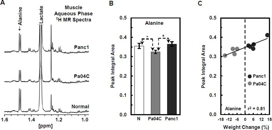
Figure 6.
1H MR spectroscopic analysis and quantification of alanine levels in skeletal muscle from normal and Panc1 and Pa04C tumor bearing mice. (A) Representative 1H MR spectra indicate a decrease of alanine in the muscle of Pa04C mice relative to Panc1 and normal mice. (B) Quantification of these differences indicates that alanine levels were significantly lower in muscle from Pa04C tumor bearing mice relative to normal and Panc1 mice (*p < 0.05 and +p < 0.1). (C) A strong positive correlation (r2 = 0.81) was found between low muscle levels of alanine and weight loss.

