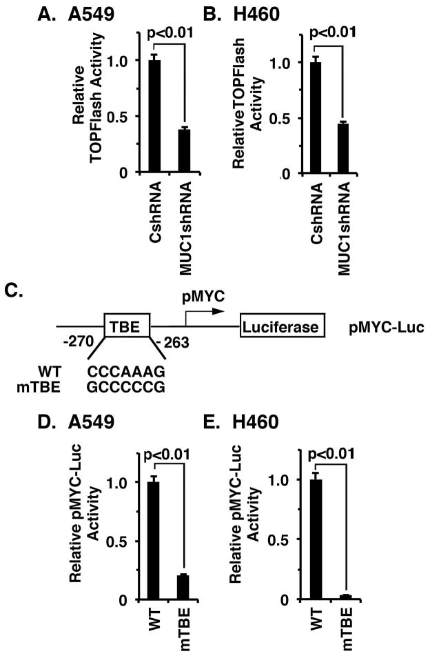
Figure 3. MUC1-C induces MYC transcription by the WNT/β-catenin/TCF4 pathway.
A and B. The indicated A549 (A) and H460 (B) cells were transfected with TOPFlash for 48 h and then assayed for luciferase activity. The results (mean±SD from 3 determinations) are expressed as relative TOPFlash activity as compared to that obtained in cells expressing the CshRNA (assigned a value of 1). C. Schema of the pMYC-Luc reporter highlighting the mutated TBE (mTBE) site. D and E. A549/CshRNA (D) and H460/CshRNA (E) cells were transfected with wild-type (WT) or TBE-mutated pMYC-Luc. The results (mean±SD of three determinations) are expressed as the relative pMYC-Luc activity compared to that for cells transfected with the WT pMYC-Luc (assigned a value of 1).

