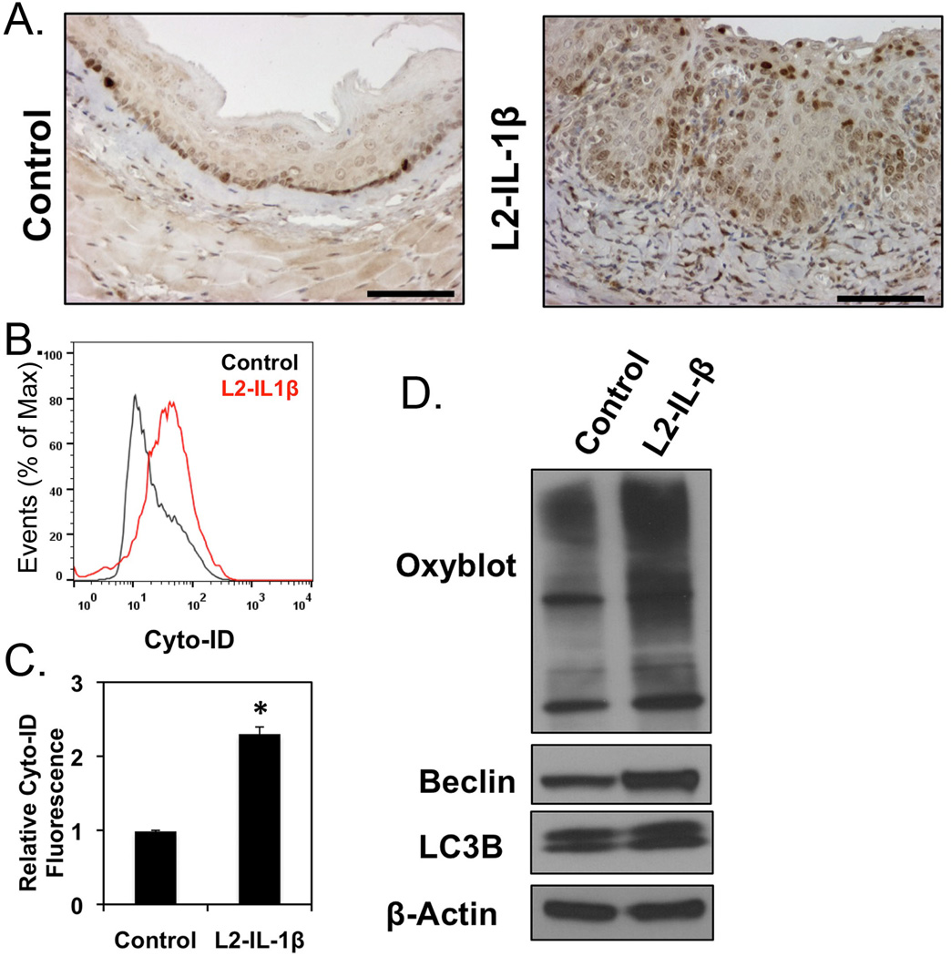
Figure 4. Oxidative stress and autophagy in the L2-IL-1β transgenic mouse model of BE.
A. IHC staining for active cleaved form of LC3 in normal mouse esophagus or transgenic L2-IL-1β mice. B. Cyto-ID autophagy profile of wild- (Black) and L2-IL-1β esophageal epithelium (Red) by flow cytometry. C. Averaged relative Cyto-ID fluorescence, n=3. *, p<0.05. D. Western blot for oxidized proteins (Oxiblot, Millipore) and autophagy proteins (LC3B and Beclin1) in the squamous epithelium from 3 month old L2-IL-1β mice. One of three blots is shown.

