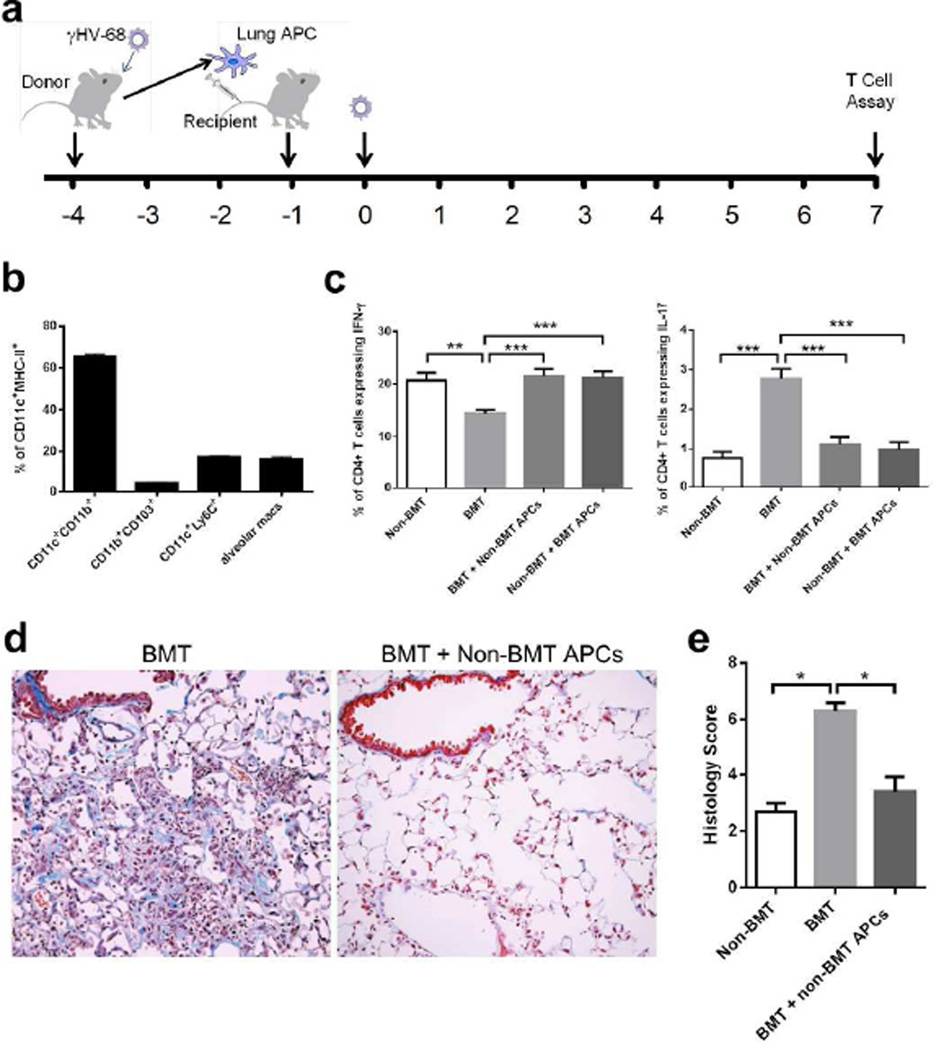
Figure 7. Lung APCs from non-BMT mice restore TH1 and limit TH17 response in BMT mice.
(a) A scheme of adoptive transfer of lung APCs and γHV-68 infection. (b) Characterization of CD11c+ MHC class II+ cell population used for adoptive transfer. (c) The alteration of the percentage of CD4+ IFN-γ+ TH1 cells and CD4+ IL-17A+ TH17 cells by adoptive transfer of lung APCs. Lung CD11c+ APCs were enriched by magnetic beads from viral-infected non-transplanted or BMT mice, and adoptively transferred into BMT mice or non-transplanted mice, respectively. Single cell suspensions were prepared at 7 dpi for PMA stimulation and flow cytometry analysis. Left, percent of CD4+ cells that express IFN-γ (TH1 cells, mean + SEM, n = 5); Right, percent of CD4+ cells that express IL-17A (TH17 cells, mean + SEM, n = 5). (d) Representative Masson’s trichrome staining on lung sections from BMT mice with or without adoptive transfer of primed non-BMT lung APCs at 21 dpi. The blue staining represents deposition of collagen. Same magnification was used for all images. (e) The average histology scores of lung sections from γHV-68 infected non-BMT, BMT mice or BMT mice adoptively transferred with non-BMT lung APCs at 21 dpi (mean + SEM, n = 3). * P <0.05, ** P <0.01, *** P <0.001. Similar results were obtained in two (b, d, e) or three (c) independent experiments.

