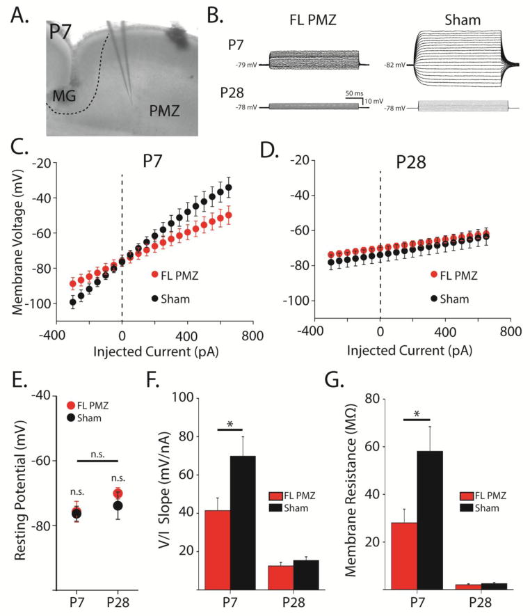
Figure 2. Astrocyte membrane resistance is decreased in the paramicrogyral zone at P7.
A, DIC image of a P7 coronal slice showing the microgyrus (MG), the paramicrogyral zone (PMZ), and the recording electrode in PMZ. B, Overlaid voltage responses in astrocytes current-clamped and injected with 20 current steps from −300 to +160 pA. C, Average voltage/current relationship for astrocytes in freeze lesion (FL) PMZ cortex (red) and sham-injured cortex (black) at P7. D, Same as (C) at P28. E, Resting membrane potentials at P7 and P28 for FL PMZ (red) and sham-injured astrocytes (black). F, Average slopes of the voltage/current relationships in (C) and (D). G, Membrane resistances of astrocytes at P7 and P28 from FL PMZ (red) and sham-injured cortex (black). Error bars represent SEM, * p < 0.05, two-sample t-test.

