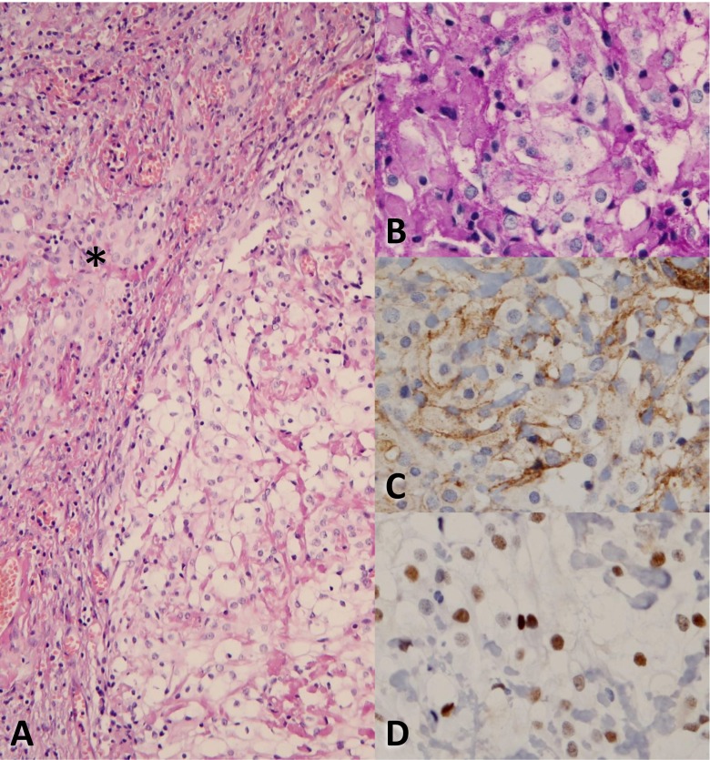
Fig. 2.
H&E staining of the tumor (a, magnification ×20), partly consisting of clear cells on the right side. The asterisk indicates meningothelial cells. In (b) to (d) more detailed micrographs (magnification ×40) of the clear cell component after PAS, EMA and progesterone receptor staining, respectively

