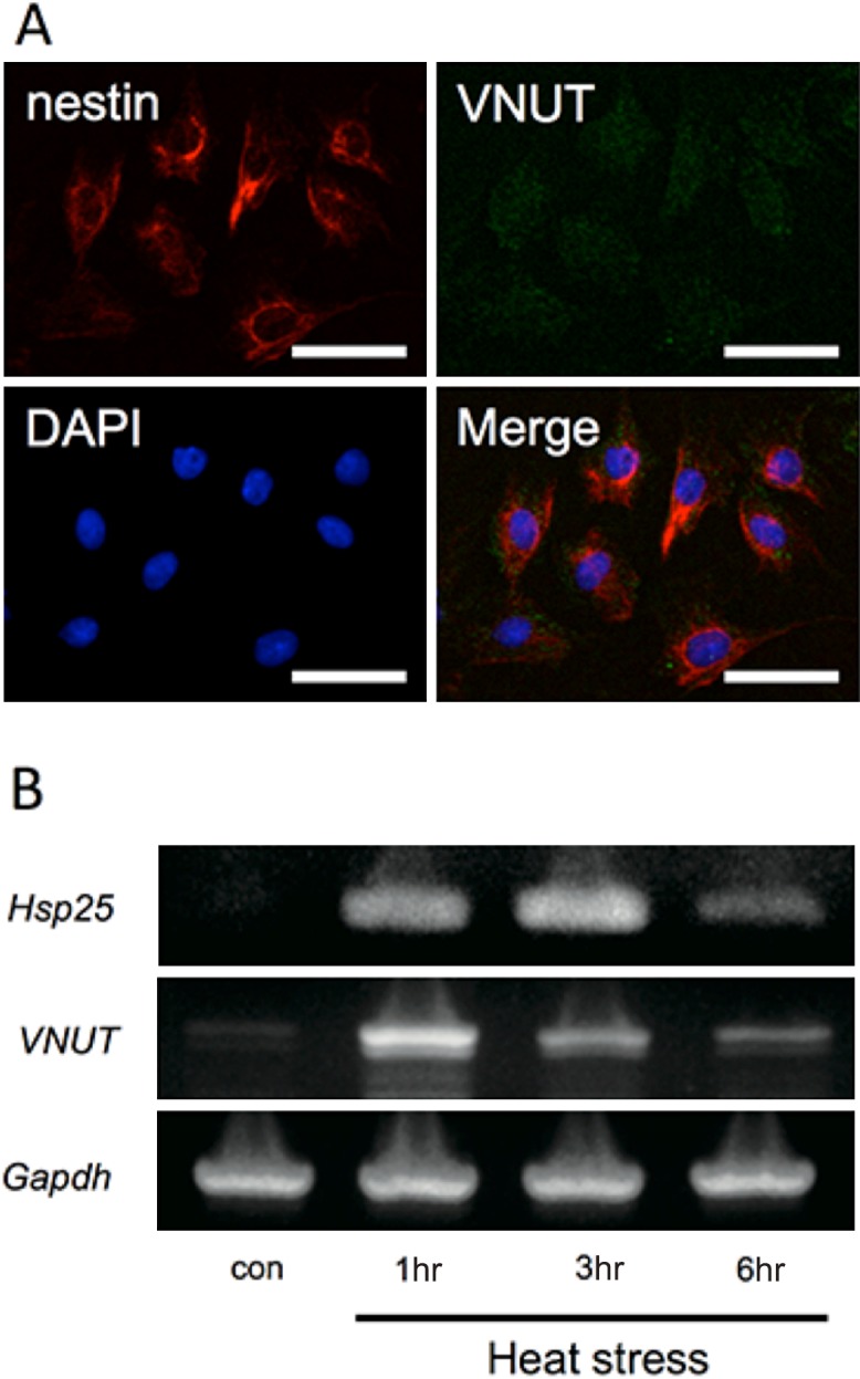
Fig. 3. .

Immunohistochemical staining in KN-3 cells and Reverse-transcriptase PCR analysis for the expression of Hsp25 and VNUT in heat-treated KN-3 cells. (A) Immunofluorescence staining for VNUT and nestin and DAPI and merged in KN-3 cells. (B) Expression of Hsp25 was observed in KN-3 cells at 1, 3, and 6 hr after heat treatment. VNUT was most strongly expressed at 1 hr after heat treatment, subsequent to which the expression decreased gradually.
